- Image A shows a stage of neurulation in a chick embryo, and image B is a scanning electron micrograph (SEM) of a transverse section through the level indicated by the line in image A.
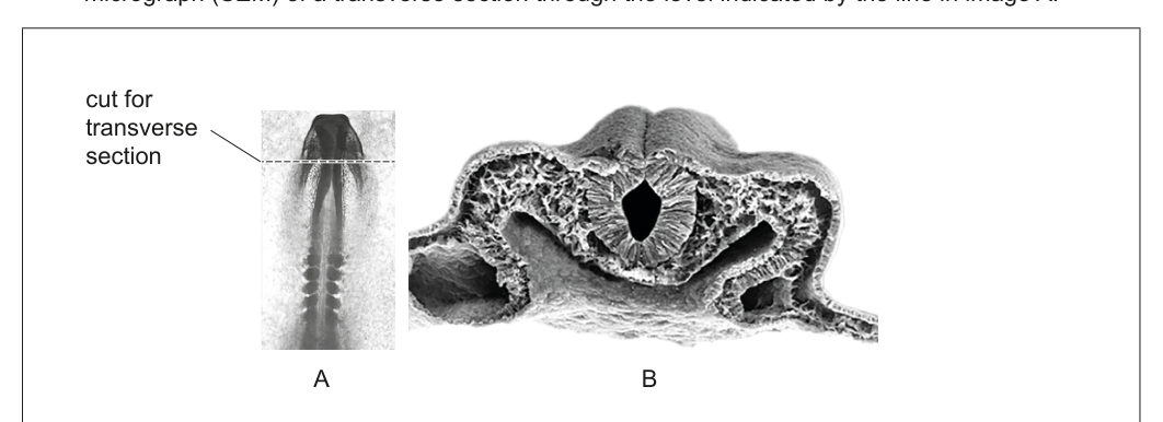
(a) On image B, label
(i) the neural tube; [1]
(ii) the ectoderm. [1]
(b) Describe the formation of neurons. [2]
(c) Explain neural plasticity. [2]